


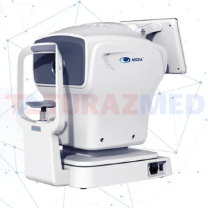
Optical ophthalmic biometer HUAS-1000
Fully automatic one-button measurement technology Click "Start measuring" button on the interface, and automatic eye-location, automatic focusing, automatic measurement can be completed. Fast measurement 3-4 groups of data collected within second, measurements for both eyes in just 25 seconds. 8 eye parameters can be obtained by one-button measurement Including axiallength, corneal curvature, axial view angle, cornea thickness, anterior chamber depth, lens thickness, white to white distance, pupil size. Eye Care Cloud The measured data can be uploaded to the eye care cloud, and the personal ophthalmic data files can be established to help prevent and control myopia in children and adolescents. Easy to operate, easy to train With only a few minutes oftraining, operators can quickly get started. Visual acuity screening for prevention and control of myopia in children and adolescents Easy operation, fast measurement, and suitable for large-scale screening projects such as schools. Establish personal visual health files for timely and effective detection of visual changes, which is suitable for prevention and control of myopia in children and adolescents. Multiple measurement modes to address requirements for different scenarios. Measurement ofaxiallength and corneal curvature helps diagnose axial myopia and refractive myopia. Specification Axial length: 12~34mm, axial resolution 0.01mm, ±25μm Corneal curvature: 4.7~11.2mm, resolution 0.01mm, ±10μm Axial view angle: 0°~180°, resolution 1°, ±9° Corneal depth: 300~800μm, resolution 1μm, ±2μm Anterior chamber depth: 1.5~6.0mm, resolution 0.01 mm, ±20μm Lens thickness: 0.5~7.0mm, resolution 0.01mm, ±50μm
Optical ophthalmic biometer MD-810A
Fully automatic one-button measurement technology Click "Start measuring" button on the interface, and automatic eye-location, automatic focusing, automatic measurement can be completed. Fast measurement 3-4 groups of data collected within second, measurements for both eyes in just 25 seconds. 8 eye parameters can be obtained by one-button measurement Including axiallength, corneal curvature, axial view angle, cornea thickness, anterior chamber depth, lens thickness, white to white distance, pupil size. Eye Care Cloud The measured data can be uploaded to the eye care cloud, and the personal ophthalmic data files can be established to help prevent and control myopia in children and adolescents. Easy to operate, easy to train With only a few minutes oftraining, operators can quickly get started. Visual acuity screening for prevention and control of myopia in children and adolescents Easy operation, fast measurement, and suitable for large-scale screening projects such as schools. Establish personal visual health files for timely and effective detection of visual changes, which is suitable for prevention and control of myopia in children and adolescents. Multiple measurement modes to address requirements for different scenarios. Measurement ofaxiallength and corneal curvature helps diagnose axial myopia and refractive myopia. Specification Axial length: 12~34mm, axial resolution 0.01mm, ±25μm Corneal curvature: 4.7~11.2mm, resolution 0.01mm, ±10μm Axial view angle: 0°~180°, resolution 1°, ±9° Corneal depth: 300~800μm, resolution 1μm, ±2μm Anterior chamber depth: 1.5~6.0mm, resolution 0.01 mm, ±20μm Lens thickness: 0.5~7.0mm, resolution 0.01mm, ±50μm

Ophthalmic perimeter MD-820A
Stimulus light source: LED; single alternating working mode; emission power no higher than 3.66mW during its operation, maintenance, repair or under faulty conditions. Stimulus light luminance: 0.5 ~ 318nt (1.6 ~ 1000asb) Stimulus light brightness level: 15 levels, each level with 2dB Brightness error of each indicator:±20%; Background light luminance: 0.02 cd/m2,±20% Dimension and shape of stimulus indicator: circular indicator with diameter of 2mm, nominal solid angle: Ω = 2.88 × 10⁻⁵, tolerance ± 15% Time characteristic for the appearance of stimulus indicator Stimulus lasting time: 0.1s, 0.2s, 0.5s selectable with tolerance of ±20% Stimulus interval time: 0.5s, 1s, 2s selectable with tolerance of ±20% Distance between chinrest center and screen center: 330mm±16mm Chinrest moving range: up-down direction ≥35mm and horizontal direction≥15mm.
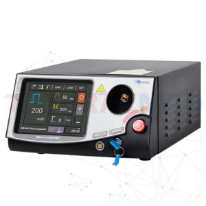
Retinal photocoagulation laser MD-960
Treatment Beam Allowable tolerance between actual output power and pre-set value is within ±15%. Pulse duration: 0.01s ~ 3s ± 5% Pulse interval: 0.05s ~ 1s ± 5%; or Single Pulse (SP) Duty cycle1: When output power is within 50mW ~ 500mW , duty cycle is not limited; Pulse Counter: 0~9,999 Laser beam spot size: 50µm~500µm, continuously adjustable, the allowable tolerance of scale is within ±15%; the allowable tolerance of minimum scale (50µm) is within ±15µm. Beam divergence angle: Adaptor output, 13.8°± 20% Repeatability of laser power: RP ≤±10 % Aiming Beam Type: Semi-conductor Laser Cooling System Primary Cooling System: Intelligent TEC (thermal-electric-coupling) semi-conductor cooling; Secondary Cooling System: Air-Cooled.
Capsulotomy laser MD-920
Laser mode: multi-mode Laser pulse output: single-pulse, dual-pulse and three-pulse Time characteristics of the laser pulse output a) Laser pulse width: 4.5ns ± 10% b) Duration of laser pulse sequence: dual-pulse≤30µs; three-pulse≤50µs c) Maximum emission repetition rate of laser pulse sequence: 2.5Hz±20% Laser output energy (in front of eyeball) a) Maximum output energy of single-pulse: 11mJ±20% b) Maximum output energy of dual-pulse: 19mJ±20% c) Maximum output energy of three-pulse: 28mJ±20% d) Output energy adjustment: 100%~6% 7-grade adjustable, the allowable tolerance between actual output and preset value is ±20% Laser output energy reproducibility: RP ≤±10% Laser beam convergence angle: 18°±20% Focal plane spot diameter: 30µm±20%
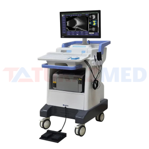
On-platform ultrasound system MD-300L
Gain: ≥105dB Digital Scan Converter Capacity: ≥1024×768×8 bit×64; Grey Scale: 256 Levels View Ports: (Width × Depth): 8.25mm×5.5mm (Narrow-view Mode) or 16.5mm×11mm (Full-view Mode) Display Delay Depth: 3mm at 16.5mm × 11mm; 7mm at 8.25mm × 5.5mm Display Mode: B Mode Applications based on Microsoft Windows XP operating system Resolution: Axial≤0.04mm; Lateral≤0.04mm Accuracy of Geometric Positions: Vertical≤10%; Horizontal≤15% Number of Image Captured: No less than 16; Video Capturing: Video saved in *.AVI format Frame-by-frame, step-by-step cineloop and dynamic cineloop Double-key Footswitch: Scan/Freeze, Image capturing and saving; Communication Interfaces: USB2.0 Interfaces Image Output: Output by a printer with USB interfaces Image Processing: Brightness and contrast adjustment, Gamma/Color Transformation, Color Conversion, Zooming and Annotation; Image Measurements: Length, Area, Angle and Perimeter Report Print-out: Chief complaints, clinical examination and imaging description printable Image File Format: JPEG, BMP Monitor Resolution: 1440 x 900 Patient Record Database Management
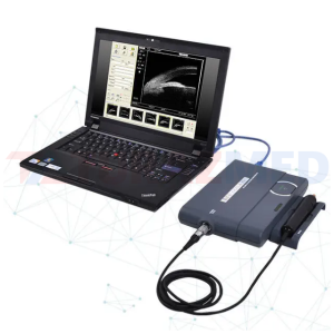
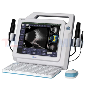
Portable ultrasound system MD-320W
Gain Control: ≥105dB Display Resolution: 1280 ×1024 or 1024 × 768 Gray Scale: 256 Delay: 3 mm - 12mm Field of View (depth × width): wide view (10.6mm × 13.5mm), normal (5.3mm ×6.7mm) Scanning Method: sector scan Resolution: Axial: 50um; lateral: 100um Accuracy of Geometrical Position: axial: ≤10%; lateral: ≤15% Display Mode: B Image Buffer Space: ≥100 images Screen Measurement: distance, angle, area and so on Communication Interface: USB Image Format: JPG, BMP Film Format: AVI Footswitch: Start\Freeze and Capture image Image Processing: Brightness and contrast adjustment, γ correction, color change, color reversal, amplification, text annotation Print Report: Observation, image content can be selected to print Hard Disk Capacity: more than 80GB Monitor Resolution: 1280 x1024
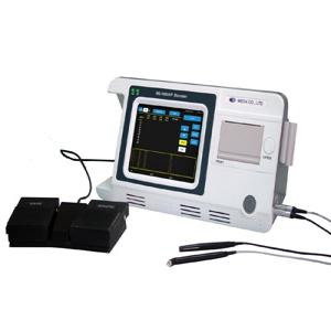
MD-1000A A Scan
The MD-1000A A scan is used to measure Axial length and calculate IOL power. With auto and manual measuring methods, it can meet the demands for different types of patients. Users may choose contact mode for quick measurement or immersion mode to obtain more accurate result. Data can be saved into the inner memory or printed out with built-in thermal printer. Uploading software kit is optional to achieve massive storage ability.

Veterinary pachymeter MD-1000P
Ultrasonic Frequency: 15-20MHz Display Resolution: 1μm Biometry Accuracy: ≤±5μm Measuring Scope: 230-1200μm

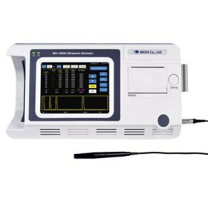
Pachymeter MD-1000A/P
A-Scan Ultrasonic frequency: 10MHz Display resolution: 0.01mm Total gain of receiver: ≥100dB Adjustable gain scope: 0~50dB Measuring scope (AL): 15 mm~40mm Measuring accuracy: ≤±0.05mm Measuring Parameter: ACD, LENS, VITR and AL Measuring mode: automatic mode or manual mode can be selectable. Automatic mode for Normal, Cataract and Aphakic mode Measuring method: contact and immersion, can be selectable IOL calculation: SRK/T, SRK-II, BINK-II, HOLLADAY, HOFFER-Q, HAIGIS, and IOL calculation formula after corneal refractive surgery: HISTORY DERIVED, DOUBLE-K/SRK-T, REFRACTION DERIVED, ROSA and SHAMMAS. Pachymeter Ultrasonic Frequency: 15-20MHz Display Resolution: 1μm Biometry Accuracy: ≤±5μm Measuring Scope: 230-1200μm
Ultrasound ophthalmic biometer MD-1000A
Ultrasonic frequency: 10MHz Display resolution: 0.01mm Total gain of receiver: ≥100dB Adjustable gain scope: 0~50dB Measuring scope (AL): 15 mm~40mm Measuring accuracy: ≤±0.05mm Measuring Parameter: ACD, LENS, VITR and AL Measuring mode: automatic mode or manual mode can be selectable. Automatic mode for Normal, Cataract and Aphakic mode; Measuring method: contact and immersion, can be selectable; IOL calculation: SRK/T, SRK-II, BINK-II, HOLLADAY, HOFFER-Q, HAIGIS and IOL calculation formula after corneal refractive surgery: HISTORY DERIVED, DOUBLE-K/SRK-T, REFRACTION DERIVED, ROSA and SHAMMAS.
ODM-2200 Ultrasonic A/B Scan
ODM-2200 Ultrasonic A/B Scanner for Ophthalmology is specially designed for doctors those having a higher demanding with his diagnosis. From the fine image to the possibility of working with PC, each feature gives a feeling of profession. Besides these, the pseudo-color enables you have a vision thrice deeper than black-white image does. While working with computer, you may save images, video and diagnostic reports as many as you like into your PC or laptop.
On-platform ultrasound system MD-2400S
B-Scan Scanning Angle: 53° Dynamic Loop: 10s/100 images, loop or frame-by-frame playback Image Saving: 100 images Gray Scale: 256 Levels Gain: 1-105dB adjustable TGC: -20dB to 20dB with dynamic range, manual sectional adjustment (6-section adjustable) Post-processing: Frame-Averaging; Pseudo-Color Codes; Gamma Correction; Text Annotation & Image Magnification A-Scan AL Biometric Measuring Range: Axial Length (AL): 15mm-40mm; AL Biometric Measuring Accuracy: No more than ±0.05mm; Total Gain: 98dB, user’s gain adjustability: 1~60dB; Measuring Mode: 5 groups (Normal, Aphakic, Special, Cataract and Manual Measurement); Measuring Method: Immersion/Contact; IOL calculation: SRK-T, SRK-II, BINK-II, HOLLADAY, HOFFER-Q and HAIGIS; Support IOL calculation after refractive surgery, two groups of formulas can be calculated contrastively and displayed simultaneously. Automatic measurement and 10 groups averaging and display standard deviation. Up to 8 groups of IOL constants can be stored.
Portable ultrasound system MD-2300S
B-Scan Scanning Angle: 53° Dynamic Loop: 10s/100 images, loop or frame-by-frame playback Image Saving: 100 images Gray Scale: 256 Levels Gain: 1-105dB adjustable TGC: -20dB to 20dB with dynamic range, manual sectional adjustment (6-section adjustable) Post-processing: Frame-Averaging; Pseudo-Color Codes; Gamma Correction; Text Annotation & Image Magnification A-Scan AL Biometric Measuring Range: Axial Length (AL): 15mm-40mm; AL Biometric Measuring Accuracy: No more than ±0.05mm; Total Gain: 98dB, user’s gain adjustability: 1~60dB; Measuring Mode: 5 groups (Normal, Aphakic, Special, Cataract and Manual Measurement); Measuring Method: Immersion/Contact; IOL calculation: SRK-T, SRK-II, BINK-II, HOLLADAY, HOFFER-Q and HAIGIS; Support IOL calculation after refractive surgery, two groups of formulas can be calculated contrastively and displayed simultaneously. Automatic measurement and 10 groups averaging and display standard deviation. Up to 8 groups of IOL constants can be stored. General Monitor: resolution≥1024×768, gray scale≥256; Keyboard and Mouse: USB interface, wireless keyboard and mouse; Output Interface: 4×USB2.0 and HDMI interface;

SNT-700 Soft Non-contact Tonometer
SNT-700 Soft Non-contact Tonometer No surprises for your patients Ensures easy examination for patients Considering safety of patients Consistent measuring accuracy Making examinations easier Easing maintenance
ARKM-200 Auto-Refkeratometer
ARKM-200 Auto-Refkeratometer New Generation of Multi-Functional Instrument


Diagnostic & Specialist MT-325UD Project...
Perimeter, the foundation of visual field testing

Diagnostic & Specialist CGT-2000 Contras...
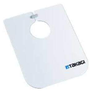
S06-71 Breath Shield
The enlarged TAKAGI breath shield designed to enhance the infection control when using slit lamps. Breath shields have been used as a physical barrier between the ophthalmologist and the patients. During the Covid-19 outbreak, TAKAGI has developed a new bigger breath shield S06-71 that provides a larger covering area than the standard size breath shield, aiming for the reduction of droplet transmission and respiratory infections.
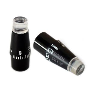
S12-18 Reusable Prism
Smart integrated structure reduces risk of infection Measuring Prism S12-18 features an integrated structure that reduces infection risk. Its environmentally conscious design is ideal for re-use, and enables highly accurate and safe intraocular pressure measurement. Methacrylic resin, one of the hardest and most highly transparent optical-use plastic materials, is used to realise brightness and clarity of observation and to product life.

Slit Lamp Microscopes AT-1 Applanation T...
R-type applanation tonometer compatible with a wide range of TAKAGI slit lamps AT-1 is based on Goldmann applanation tonometry and designed for use with TAKAGI slit lamps, the AT-1 (R-type) provides rapid, safe, and accurate intraocular pressure (IOP) measurement required for the diagnosis of such conditions as glaucoma, ocular hypertension, and retinal detachment.